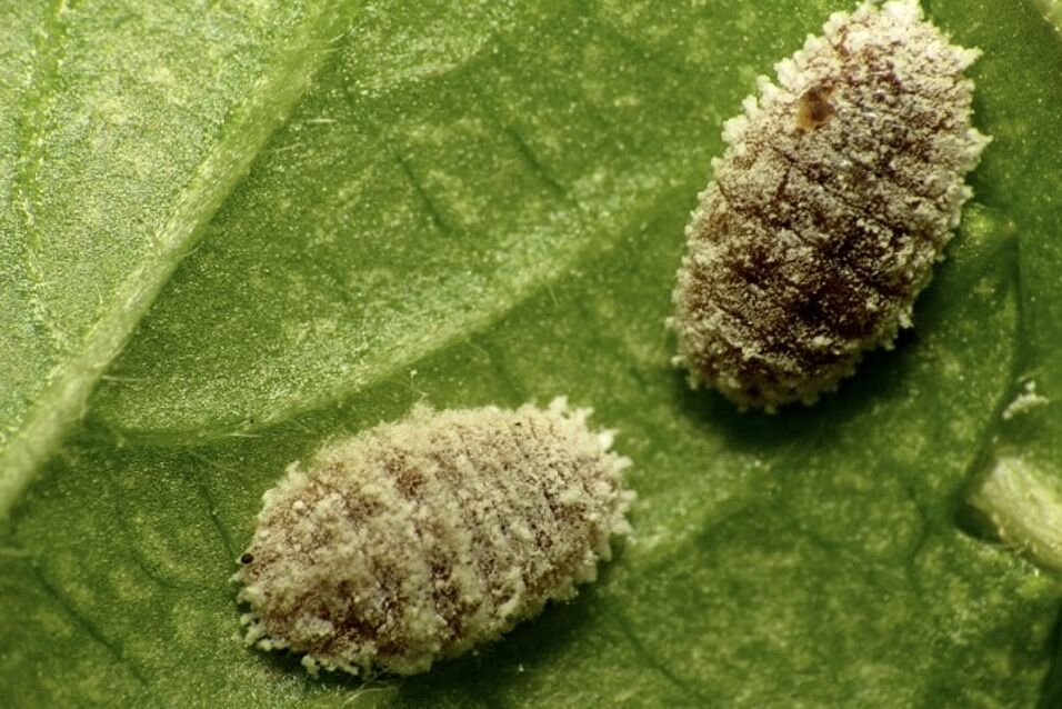
We are currently studying the bacterial symbionts of two insect groups, adelgids (Adelgidae) and mealybugs (Pseudococcidae). The symbionts of mealybugs show a unique feature which makes this symbiosis different from all other known symbioses: The two phylogenetically different bacterial symbionts of mealybugs are not located in the same or neighboring bacteriocytes, but one symbiont (Tremblaya princeps) contains the other symbiont, which lives directly within its cytoplasm. Our work will contribute to a better understanding of this nested symbiosis and the biology of a group of mealybugs which occurs as a major pest all over the world. It also addresses fundamental questions such as the establishment and evolution of endobacterial symbiosis, which has implications for our understanding of early evolutionary processes leading to the first eukaryotic cell.
The adelgids are a sister group of aphids (Aphididae), the probably best studied groups of insects with respect to bacterial symbionts. Adelgids are plant sap feeding insects which exclusively feed on conifers, and whose symbionts are only poorly characterized so far. We found that adelgids contain a surprising diversity of bacterial symbionts suggesting a complex evolutionary history for this symbiosis, which involved co-evolution as well as multiple infection events and symbiont replacement. This is fundamentally different from symbiosis between bacteria and aphids, where a single obligate symbiont, B. aphidicola, is present in nearly all aphid species since more than 150 million years. The investigation of additional adelgid species and genomic analysis of the identified symbionts will help to further extend our understanding of the evolution of this symbiosis and the role of the bacterial symbionts in this association.
This project is funded by the Austrian Science Fund (P22533-B17).

