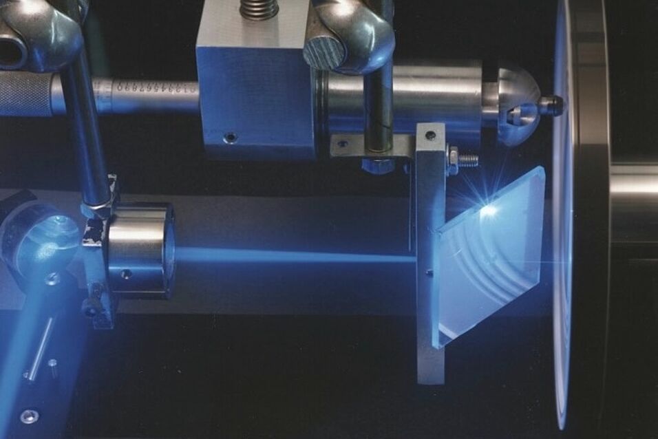
Raman microspectroscopy is a powerful technique for the analysis of microbial cells at the single cell level. A single Raman spectrum of a microbial cell can be acquired within seconds and yields data about its abundant chemical bonds, such as those derived from nucleic acids, proteins, lipids, and carbohydrates. Raman spectra provide chemical fingerprints that are highly sensitive to the cellular biochemical composition and physiological state of the cell. Raman also allows substrate utilization to be monitored: by providing 13C labelled variants of compounds of interest, assimilation of 13C into cellular biomass can be detected by distinctive shifts in peaks in the Raman spectrum. Raman microspectroscopy is non-destructive, and the activity of organisms can be monitored over time without affecting their physiology.
In this project, a novel microcosm-imaging platform will be established and used to visualize interactions in realistic, heterogeneous, three-dimensional environments. This will serve as a robust system to manipulate, perturb, and observe microbial activity in situ that is also flexible enough for diverse applications by the broader scientific community. This approach will improve our understanding of how complex microbial communities are generated, maintained, process substrates, and may be usefully manipulated.
This project is funded by the U.S. Department of Energy.
Investigated by:
- Berry D
- Palatinszky M
- Nikolov G

